The Test
Congenital heart disease is a leading cause of infant morbidity and mortality from birth defects with an estimated incidence of 6 (8 to 9) per 1000 live births for moderate to severe forms. Most cases are not associated with known risk factors.
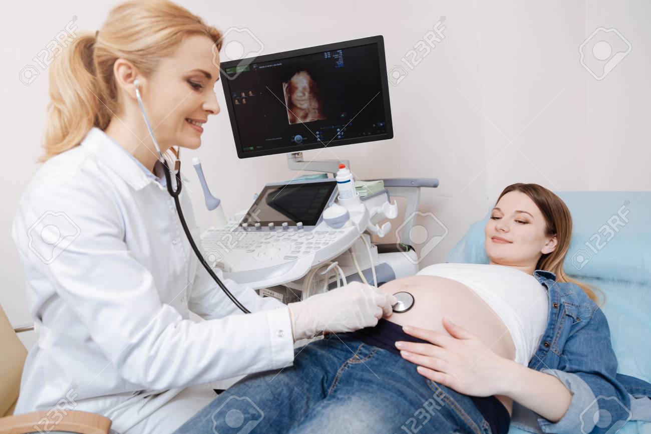
Fetal echocardiography is broadly defined as a detailed sonographic evaluation that is used to identify and characterize fetal heart anomalies before delivery.
The test is typically performed by specially trained Fetal Medicine Specialists.
A limited evaluation of the fetal heart is possible during regular obstetric scanning. However, Fetal Echocardiography is an important tool in detailed assessment of fetal heart, which is certainly recommended in many At Risk Pregnancy situations.
In Routine Low Risk Pregnancies, it can be an optional test to reasonably rule our major cardiac defect in the baby. Ultrasonography through maternal abdomen is the most common method used to evaluate the baby’s heart. However, rarely Trans-vaginal Route (TVS) may be necessary.
The Time
Fetal echocardiograms can be performed after 18 weeks gestation. However, if someone had previous normal Fetal Anatomy Scan, Fetal Echo is usually performed around 22-24 weeks.
The Result
At the end of the assessment, you will be told if no fetal cardiac problems are found. But, it is important to know that even in the case of a totally normal examination, not every heart problem can be ruled out by examination inside uterus. This is because the circulation in the fetus is different than after birth. Additionally, very small holes between the lower chambers of the heart are hard to see. However, considering the normal fetal circulation, your doctor can provide fairly definitive good news in the case of a normal fetal echocardiogram.
If a heart defect is found, you will be referred to a Paediatric Cardiologist for more detailed diagnosis and counselling regarding outcome, treatment and the need for heart surgery after birth.
Points to Note
- This test is not painful and causes no harm to the baby.
- The test can take longer than other Fetal Scans, depending on fetal position, pregnancy gestation, maternal body weight and complexity of the baby’s heart problems, if any. A full bladder is not necessary for a fetal echocardiogram.
- It is always important to have as much information as possible when you come for your fetal echocardiogram, especially the details of why you were referred by your obstetrician, like any medical history in mother, any history of previous baby with heart problems. A four fold increase in congenital heart diseases is noted in fetuses of IVF pregnancies.
- There also may be a structure that is not seen as well as the doctor would like, and you may be asked to return even though the suspicion of a problem is low. Some problems such as maternal lupus may require more than one study even if the first one is normal.
- Certain heart defects may significantly increase the risk of genetic problems such as Down Syndrome or DiGeorge Syndrome. The finding of benign tumours in the heart makes the diagnosis of Tuberous Sclerosis, a genetic syndrome that has significant implications for abnormal brain development much more likely. These issues may have significant implications on the prognosis of the child and play a major role in helping you make decisions about your pregnancy. Thus, sometimes when a congenital heart disease is found, a genetic assessment of the baby may be needed, either after delivery or before by invasive testing like Amniocentesis.
The Interpretation
- Based on the calculation, the test will be reported as either Negative Screening (Normal result) or Positive Screening (Abnormal result). Parents will receive full counselling concerning the significance of the positive test and the various options for further testing .Only you can then decide if you wish to have an invasive diagnostic test for final confirmation of any problem in baby.
- If the test is reported as normal, it is recommended that you have an Anomaly Scan at 20 weeks to check for physical abnormalities in the baby.
The Preparation and After
- You may eat normally before.
- We will need a reasonably full bladder for the test so that you need to drink sufficient water for about an hour before the test and hold your urine.
- After the sonography is done, you will be taken for a blood sample collection in the normal way after which you may leave. Result should be available in about 5 working days